Figure 1.

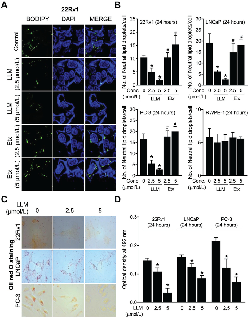
Leelamine (LLM) treatment decreased number of neutral lipid droplets in prostate cancer cells. A, Representative confocal microscopy images for BODIPY staining depicting neutral lipid droplets in 22Rv1 cells after 24-hour treatment with ethanol (control) or 2.5 and 5 µmol/L of LLM or Etomoxir (Etx; positive control). B, Quantitation of the number of neutral lipid droplets/cell in control and LLM-treated 22Rv1, LNCaP, PC-3, and RWPE-1 cells (24-hour treatment). Results shown are mean ± SD (n=3). Significantly different (P<0.05) *compared with ethanol-treated control, and #between LLM and Etx group by one-way ANOVA followed by Bonferroni’s test. C, Oil red O staining in 22Rv1, LNCaP, and PC-3 cells after 24 hours of treatment with ethanol (control) or the indicated doses of LLM. D, Quantitation of Oil Red O colour intensity through measurement of absorbance at 492 nm. Results are shown as mean ± SD (n=3). *Significantly different (P<0.05) compared with corresponding ethanol-treated control cells by one-way ANOVA followed by Dunnett’s test. Consistent results were observed in independent replicate experiments.
