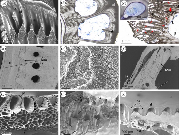
Figure 2.
Main morphological elements in the Balanidae. (a) View of the basal growth margin of a paries of P. perforatus. (b) Transversal section through a paries of A. psittacus. Growth lines indicate growth of the outer lamina towards the interior. The inner lamina coarsens both towards the interior and the exterior and incorporates the denticulate parts of the primary septa (see panel (a)) as interlaminate figures. Note cellular material within the canals. (c) Longitudinal view of the base and wall plates of A. psittacus. The thin red arrows point to sectioned tubes of the base plate and other void structures at the contact of the base plate with the wall plates, which contain remains of cellular tissue. The thick red arrow points to the initiation of a radius. The inset is a transversal section through a longitudinal canal showing the aspect of the infilling tissue. (d) View of a horizontally sectioned paries of P. perforatus, showing internal organic membranes. (e) Fibrous membrane within a lateral plate of P. perforatus, partly exhumed by slight decalcification. (f) Horizontal section through the contact between plates of P. perforatus, showing the distribution of internal membranes; they are particularly patchy in the radius region. (g) Fracture of the margin of the base plate of A. psittacus, showing the difference between the primary and subsidiary tubes. Note the small grooves accommodating the denticles of the primary septa at the initiation of the primary tubes (thick arrows). Remains of two such septa show the existence of permanent interstices between the denticles and the primary tube wall (thin arrows). (h) Partly decalcified specimen of A. psittacus, showing the interlocking between the primary septa of the paries base and the ends of the primary tubes of the base plates. Decalcification reveals the organic linings of the primary and subsidiary tubes. (i) Interlocking between the denticulate portions of the primary septa and the primary tubes in A. psittacus. Note permanent gaps between the inner lamina and the basal plate. (a), and (d) to (i) are SEM views; (b) and (c) are optical microscope views. a, ala; BP, basal plate; CM, carinomarginal plate; iaf, intralaminate figure; if, interlaminate figure; il, inner lamina; iom, internal organic membrane; la, longitudinal abutment; ol, outer lamina; p, paries; ps, primary septum; pt, primary tube; R, rostrum, r, radius; RM, rostromarginal plate; ss, secondary septum; st, subsidiary tube; tl, translucent layer.

