Figure 3.
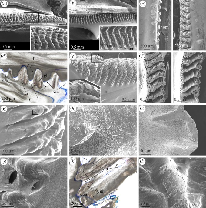
Structural elements found at the lateral boundaries between plates. (a) View of the carinal margin of the radius of a wall plate of P. perforatus. The inset is a detail of the crenulations, showing their dendritic margins. (b) Rostral margin of the paries directly opposing the specimen in (a). The inset shows the system of crests and troughs covered by an organic membrane. (c) Profile views of the rostral margin of the paries (left) and the opposing carinal margin of the radius (right) in P. perforatus. Note the membrane covering the paries margin and the much higher elevation of the crenulations of the radius margin. (d) Cross-sectional view of the contact between the carinal margin of the radius and the rostral margin of the paries in A. psittacus. There are permanent voids between the troughs of the former margin and the crenulations of the latter. Some of them show remains of cellular material. The crenulations of both margins display internal growth lines. (e) Aspect of the carinal margin of the radius of a wall plate of A. psittacus, close to the plate base. Note the particularly dendritic aspect of the crenulations. The division into lower-order branches takes place towards the plate interior. The inset shows the smooth texture of the crenulation surfaces. (f) Different designs of carinal margins of radii of two specimens of A. psittacus. The division into new high-order branches takes places towards the shell interior (towards the right of images). (g) Incipient crenulations in A. psittacus. Dendrites begin to form within each crenulation with the development of gully-like structures. The texture of the top areas of the crenulations is particularly smooth. (h) Detail of the area framed in (g), showing the difference in texture and grain size between the top area of the crenulation and the interior of one of the gullies. (i) Detail of the edge of a crenulation of A. psittacus, showing the difference in texture between the flat top and the slope. (j) System of crests and troughs of the rostral margin of the paries of A. psittacus. Their surfaces are smooth and the intervening intermediate membrane is partly detached from the surface. (k) Horizontal section across the contact between the radius and paries in A. psittacus. The radius bears internal growth lines. The intermediate organic membrane is stained in blue. Note the existence of cellular tissue filling in the voids left at the troughs of the crenulations of the radius margin. (l) View of an area similar to that in (j), in P. perforatus. The intermediate organic membrane is partly abraded; however, its smooth texture is still evident. (a) to (c), (e) to (j) and (l) are SEM views; (d) and (k) are optical microscope views. cm, cellular material; cr, crenulation; gl, growth line; om, organic membrane; p, paries; r, radius; v, void.

