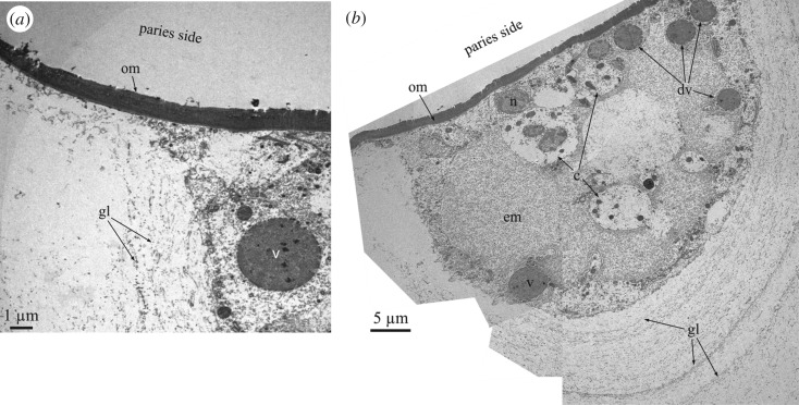
Figure 4.
TEM analysis of the membranes observed at the radius–paries boundaries and associated cellular material. (a) View of the organic membrane between the carinal margin of the radius and the rostral margin of the paries of A. psittacus. (b) Composite micrograph of the cellular material found at the trough between crenulations of the carinal margin of the radius of the wall plate of A. psittacus (cf. figure 3d). Upon decalcification, the growth lines are revealed by aligned fibrils. c, cell; dv, v, dense vesicles; em, extracellular matrix; gl, growth lines; n, cell nucleus; om, organic membrane.

