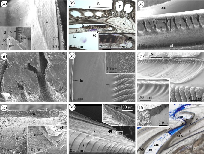
Figure 5.
Structural elements found at the contacts between the alae and sheaths. (a) General view of the sheath–ala complex of A. psittacus. The crenulated margin of the ala is separated by a cleft. The inset is a fracture through the ala and the sheath of the opposite plate, showing the mode of insertion of the ala into the lateral abutment. (b) Horizontal cross section through the junction between the ala of the wall plate and the sheath of the rostrum in A. psittacus. The ala and sheath of the wall plate can easily be differentiated by the presence of growth lines in the ala. The inset is a detail of the junction, in which the intermediate organic membrane (stained in blue) can be discerned. (c) Crenulated margin of the ala in A. psittacus. Note the incipiently dendritic aspect of the crenulations. The intermediate membrane can be seen at the top. The cleft separating the crenulated edge from the rest of the ala is at the bottom. (d) Detail of the area framed in (c). The top areas of the dendrites have a smooth surface texture. (e) View of the articular surface for the adjacent ala and the radius edge of a wall plate of A. psittacus. The inset is a detail of the framed area and shows the difference in surface texture between the articular surface and the rest of the internal surface of the plate. (f) Similar view in P. perforatus. The inset is a detail of the framed area and shows the set of smooth crests and troughs of the base of the longitudinal abutment, in which the edge of the ala fits. (g) Aspect of the organic membrane covering the ala edge in A. psittacus. The inset is a detail of the same type of membrane in P. perforatus. Its surface is imprinted with growth lines. Note the fine-grained texture of the crenulation surface. (h) Aspect of the sheath–ala complex in P. perforatus. The arrow marks the boundary between the denticulate and non-denticulate sections of the edge of the ala. The inset is a view of the non-denticulate part of the edge of the ala (framed area). (i) Horizontal section through the contact of ala and sheath in A. psittacus. The intermediate membrane between the edge of the ala and the longitudinal abutment appears stained in blue. This membrane continues into a thick and dense laminated connective organic structure, which extends in the direction towards the radius edge. The cellular material responsible for the secretion of this thick membrane is indicated. The inset is a TEM view of the contact between the connective organic structure and the secreting cells, taken from an area similar to that framed in the optical micrograph. (a) and (c) to (h) are SEM views; (b) and (i) are optical microscope views. a, ala; cl, cleft of the alar margin; cm, cellular material; cos, connective organic structure; gl, growth line; la, longitudinal abutment; om, organic membrane; p, paries; R, rostrum; r, radius; RM, rostromarginal plate; s, sheath; se, sheath extension.

