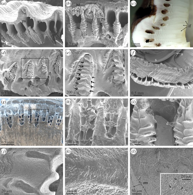
Figure 7.
Structural elements observed at the contacts between the wall plates and the basal plate. (a) View of the base of a rostral plate of A. psittacus, showing the system of primary septa and canals. (b) Similar view of a wall plate of P. perforatus. Note the presence of abnormal outgrowths with dendritic outlines. (c) Polished section of the contact between the rostromarginal and carinomarginal plates of A. psittacus. The interlaminate figures are particularly extensive and there is one (arrow) which is denticulate all along its extension. (d) Peripheral area of the base plate of P. perforatus. Remains of the denticulate parts of the primary septa, broken off upon detachment, remained in their original position at the ends of the primary tubes of the base plate. (e) Detail of the area framed in (d). The arrows point to permanent interstices between the denticles of the primary septa and the walls of the primary tubes. (f) View of the contact between the basal plate and carinomarginal plate in A. psittacus. (g) Peripheral area of the basal plate of a juvenile A. psittacus. Upon drying and detachment, the organic membranes at the ends of the primary tubes of the basal plate replicated the outlines of the primary septa, including the denticles, which were formerly in contact with them. The back scatter mode allows us to discern the areas which contain calcite (in white) from those purely organic (in dark hue). (h) Detail of the area framed in (g) showing the imprints of the denticles of the primary septa. (i) Aspect of the denticulate parts of the primary septa in P. perforatus. The edges of the septa and denticles have a different texture from the rest of the septa. (j) Detail of the edges of denticles of primary septa. Here, their smooth texture can be appreciated. (k) Close-up view of one such edge. Apart from the smoother surface texture, the grain size is much smaller than that of the intermediate areas. (l) Polished and etched section through an interlaminate figure in P. perforatus. The branches have been outlined with white broken lines. The inset shows that the branches are made of particularly small grains; arrows point to branches. All SEM views, except for (c) (optical micrograph, reflected light). a, ala; ao, abnormal outgrowth; d, denticles; if, interlaminate figure; ps, primary septum; s, sheath; ss, secondary septum; st, subsidiary tube.

