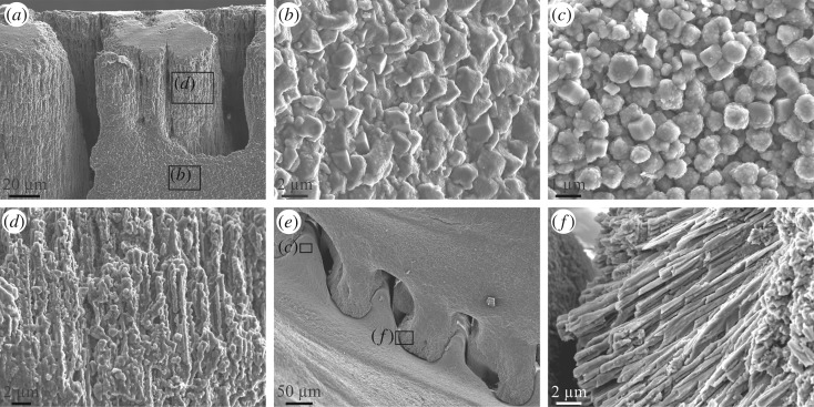
Figure 8.
Microstructures observed in A. psittacus. (a) View of the alar margin of a rostromarginal plate. Whereas the crenulations are made of calcite fibres, the shell interior has a granular rhombohedral microstructure. (b) Calcite granules observed in the wall of the ala shown in (a) (framed area). (c) Calcite granules (mostly rhombohedra) observed in the internal surface of a crenulation of the paries shown in (e) (framed area). (d) Close-up view of the fibrous microstructure of the alar crenulation shown in (a) (framed area). (e) View of the interlocking system of the crenulations of a radius (top of the image) and the opposing paries (bottom of image). Note the fibrous nature of the radius crenulations (f) and the granular nature of the paries crenulations (c). (f) Detail of the fibres observed within the radius crenulation shown in (e) (framed area). All SEM views.

