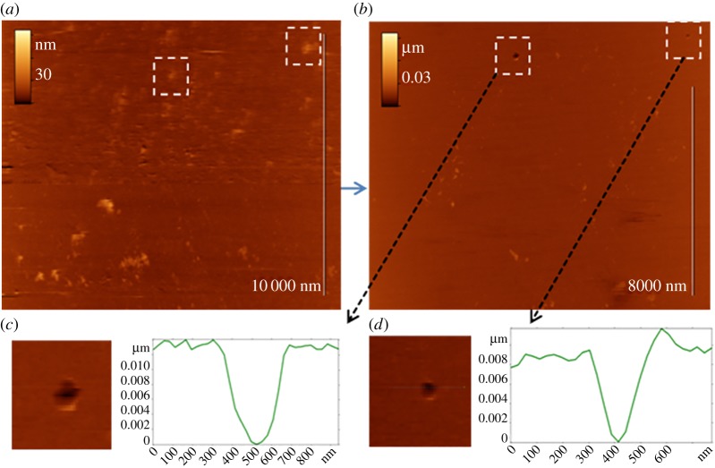
Figure 7.
AFM images of sensor layer substrate after virus adsorption. (a) Virus particles are partially dipped in the receptor layer; (b) virus particles are removed by force and applied by AFM cantilever. Marked areas on AFM images show places with partially dipped virus before (a) and holes after (b) force application; (c,d) enlarged images and cross sections of remaining holes. Scan size 11 × 14 µm2, insets 1 × 1 µm2.

