Figure 2.
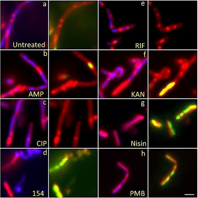
Fluorescent microscopy images of B. subtilis 6051 treated with DAPI (Blue), SytoxG (Green) and Fm4-64 (Red) treated with traditional antibiotics influencing each biosynthetic class (DNA synthesis, RNA synthesis, cell wall synthesis, protein synthesis, and membrane-active) and test compound DPA 154, suspected of being a DNA synthesis inhibitor by way of topoisomerase 1A inhibition. DPA 154 produces lysed cell contents similar to membrane-active compounds and DNA morphology similar to ciprofloxacin. Ampicillin produces filamentous cells with membrane blebbing and membrane permeation. Ciprofloxacin produces filamentous cells with reduced DNA content as seen by DAPI and a lack of permeated membranes. Rifampicin produces moderate cell lengthening and unilateral membrane bulging seen as dense membrane enhancement by FM4-64. Kanamycin procures peptidoglycan thickening, bulging, and moderate filamentation. Nisin and polymyxin B both cause cell lysis and production of ovoid cells due to compromised cell membrane. Bacteria shown in paired images; DAPI + FM4-64 (left) and FM4-64 + SytoxG (right). (a) Untreated cells. Treatment with (b) ampicillin at five times MIC (1 µg/mL), (c) ciprofloxacin at five times MIC (0.025 µg/mL), (d) 154 at 10 µg/mL, (e) rifampicin at five times MIC (0.025 µg/mL), (f) kanamycin at five times MIC (10 µg/mL), (g) Nisin at five times MIC (10 µg/mL), and (h) polymyxin B at five times MIC (5 µg/mL). Further description of expected phenotypes may be found in Supplementary Data (Tables S1–S6). Large composite images of each antibiotic may also be viewed in Supplementary Data (Figs S5–S18). Scale bar 1 µM.

