Figure 5.
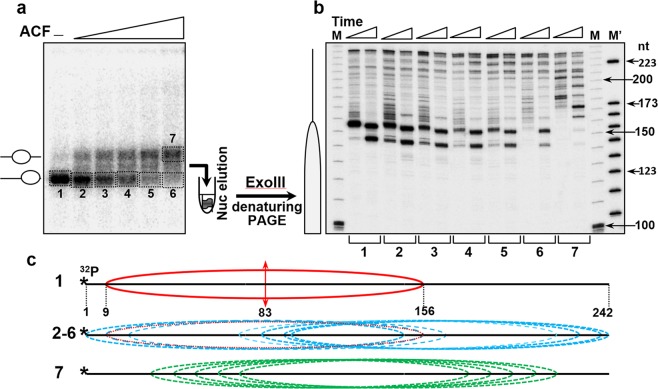
Exonuclease III mapping. (a) End-positioned nucleosomes were reconstituted on a32 P-labeled 242 bp 601 DNA sequence and incubated with increasing amounts of ACF. The reaction mixtures were then run on a 5% PAGE under native conditions, the bands corresponding to the higher mobility fractions (1–6) and to the lower mobility one (fraction 7) were excised from the gel and eluted. The gel eluted particles were next digested with 0.08 u/μl of Exo III for 1 and 2 minutes at 37 °C and the DNA was then purified (see schematics at the left part of the panel). (b) Determination of the nucleosome positions in the ACF remodeled particles. DNA, purified from the remodeled by ACF and Exo III cleaved particles was run on 8% PAGE under denaturing conditions. (M) and (M’), DNA size markers. The lengths of some of the markers (in nucleotides, nt) are noted on the right part of the figure. Left, schematic presentation of the position of the control, end-positioned nucleosome. (c) Schematics of the positions (relative to the DNA ends) of the control particles, not treated with ACF (1), the ACF remodeled particles with no changes in their electrophoretic mobility (fractions 2–6), and the particles with lower mobility (7). The single position of the control particle is shown in red, the positions of the mobilized particles from fractions 2–6 are in blue, and those within fraction 7 are in green.

