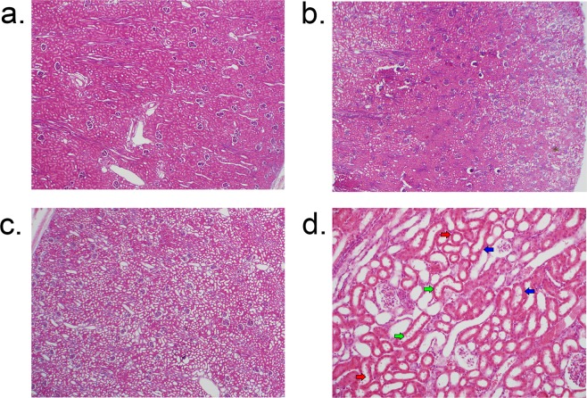
Figure 1.
Hematoxylin-eosin staining of renal cortex for acute kidney injury histology scoring. (a) Representative section from an anesthesia control animal demonstrating normal histology without evidence of AKI on low magnification (4x); (b) Section from an animal 4 hours after CPB/DHCA demonstrating moderate AKI with 25–50% of cortical area affected; (c) Section from an animal four hours after CPB/DHCA demonstrating severe AKI with 75–100% of cortical area affected; (d) high-magnification (20x) view of damaged proximal tubules demonstrating proximal tubular dilation (red arrows), epithelial thinning (blue arrows), and epithelial blebs (green arrows). CPB/DHCA = cardiopulmonary bypass with deep hypothermic circulatory arrest; AKI = acute kidney injury.

