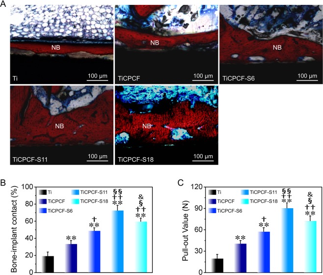
Figure 8.
(A) Histological analysis of the bone/implant interface after implantation into the rabbits for 8 weeks. Red-stained tissues represent newly formed bones. Bone-to-implant contact (B) and pull-out force (C) of the metallic Ti wires with and without coatings 8 weeks after implantation. All values are averages calculated from four independent measurements. Errors are standard deviations. **p < 0.01 compared to metallic Ti substrate; †p < 0.05, ††p < 0.01 and †††p < 0.001 compared to TiCPCF coating; §p < 0.05, §§p < 0.01 compared to TiCPCF-S6 coating; &p < 0.05 compared to TiCPCF-S11 coating.

