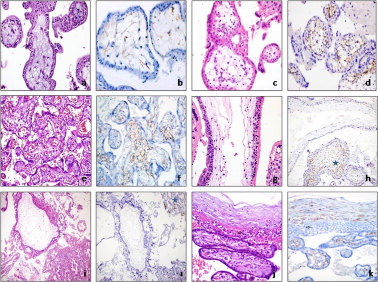
Figure 1.
(a,b) Group 1: Avascular mesenchymal villi show weak focal podoplanin immunoreactivity (a, hematoxylin and eosin [H&E], ×400; b, podoplanin, ×400). (c,d) Group 2: Significant podoplanin immunoreactivity from the sub-trophoblastic area in secondtrimester placenta, in which fetal vessels are present (c, H&E, × 400; d, podoplanin, ×400). (e,f) Group 3: Diffuse podoplanin immunoreactivity in term placental tissue (e, H&E, ×200; f, podoplanin, ×200). Group 5: Molar pregnancy groups. (g,h) In partial moles, podoplanin loss was observed in cistern-containing villi and podoplanin expression was maintained in normal appearing villi [asterisk] (g, H&E, ×630; h, podoplanin, ×400). (i,I) Total podoplanin loss of hydropic villi in a case of complete molar pregnancy. In the gestational endometrium (internal positive control), the lymphatic endothelium is positive for podoplanin [asterisk] (i, H&E, ×200; i, podoplanin, ×200). Group 6: In cases of chorioamnionitis, podoplanin immunoreactivity in chorionic villi stromal cells was similar to that in the control group (arrow indicates acute inflammation) (group 3) (j, H&E, ×400; k, podoplanin, ×400).

