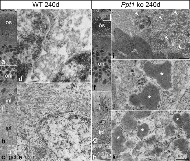
Figure 3.
Ultrastructural analysis of storage material in the retina of Ppt1 ko mice. Low-power electron micrographs depict the different layers of a 240-day-old wild-type (a–c) and age-matched Ppt1 ko retina (f–h). Analyses of the boxed areas in (f–h) at higher magnification revealed the presence of granular osmiophilic deposits in subretinally located macrophages (arrowheads in i), retinal interneurons (asterisks in j) and retinal ganglion cells (asterisks in k) respectively of Ppt1 ko retinas. Similar cytoplasmic inclusions were not detectable in retinas of age-matched control mice (d,e). gcl: ganglion cell layer; inl: inner nuclear layer; ipl: inner plexiform layer; onl: outer nuclear layer; os: outer segments. Scale bar in (h) (for a–c,f–h): 5 μm; in (i): 1 μm; in (k) (for d,e,j,k): 0.2 μm.

