Figure 8.
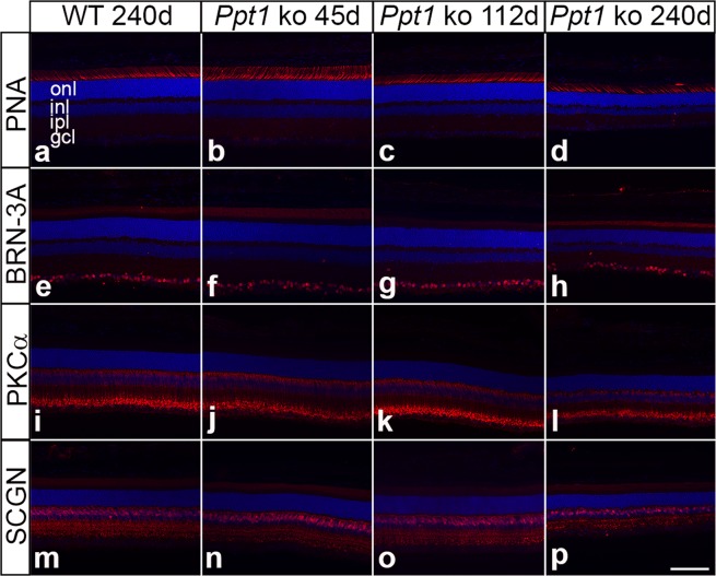
Degeneration of various retinal cell types in PPT1-deficient mice. The density of PNA-labelled cone photoreceptor cells, BRN-3A-positive retinal ganglion cells, PKCα-positive rod bipolar cells and SCGN-positive cone bipolar cells in 45-day-old Ppt1 ko retinas (b,f,j,n, respectively) was similar to that observed in wild-type retinas (a,e,i,m, respectively). Retinas from 112-day-old Ppt1 ko mice also contained normal numbers of retinal ganglion cells (g) and rod bipolar cells (k), but decreased densities of cone photoreceptor cells (c) and cone bipolar cells (o). Note the significant decrease in the number of all cell types in 240-day-old mutant retinas (d,h,l,p) when compared to age-matched wild-type retinas (a,e,i,m). gcl: ganglion cell layer; inl: inner nuclear layer; ipl: inner plexiform layer; onl: outer nuclear layer. Scale bar in (p) for (a–p): 100 µm.

