Fig. 3.

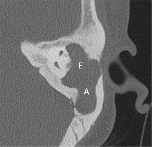
Axial CT reveals widening of aditus (denoted by A) and formation of common cavity between epitympanum (denoted by E) and aditus with soft tissue within

Axial CT reveals widening of aditus (denoted by A) and formation of common cavity between epitympanum (denoted by E) and aditus with soft tissue within