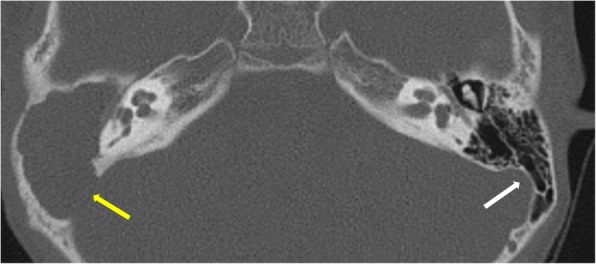
Fig. 7.

Axial CT shows normal “ice cream cone” configuration of ossicles in epitympanum on the left side formed by the head of the malleus and the body of the incus. The right ear shows soft tissue in the middle ear and aditus with non-visualization of ice cream cone suggesting erosion. A large defect is also seen in the sinodural plate on the right side (yellow arrow). Left sinodural plate is intact (white arrow)
