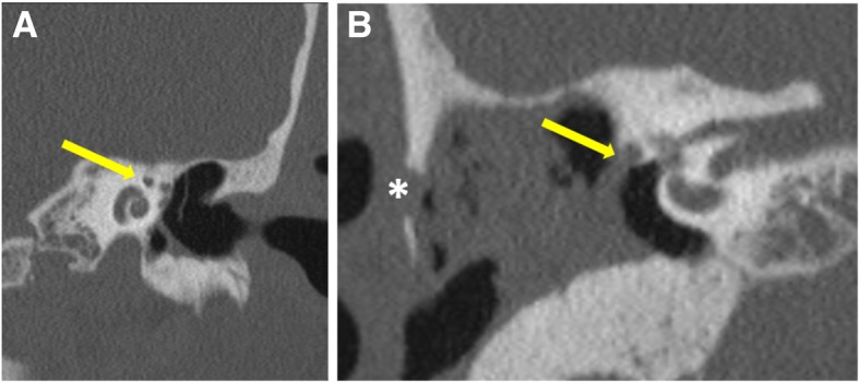
Fig. 9.

Coronal (a) CT image reveals the “snail eye” view where the turns of cochlea form the body of snail and labyrinthine and tympanic segments of facial nerve being the eyes (arrow). Coronal (b) CT image shows the erosion of the floor of the horizontal segment of the facial canal (arrow). Erosion of lateral mastoid cortex is also seen (asterisk)
