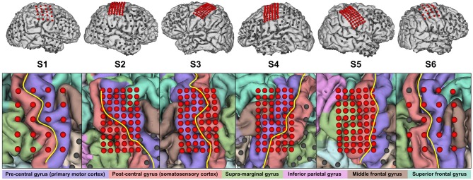
Figure 1.
Electrode placement overview. Electrodes reported in Table 2 are highlighted in red. Not all of the remaining electrodes in the top row are visible due to occlusion. In the close-up view, the central sulcus is indicated in yellow and the identified gyri are shaded in respective colors.

