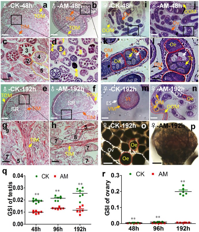
Figure 2.
Impact of hyperproteinemia on gonad development. (a-n) Sections of gonads stained by hematoxylin-eosin at 48 and 192 h after modeling. (o, p) Ovaries under cover glass at 192 h after modeling. TOM, outer testis membrane; TIM, inner testis membrane; SR, spermatogenic room. Numbers 1-3 indicate early developmental stage spermatocysts during the 32-, 64-, and 128-primary spermatocyte stages, respectively; 4 and 5 indicate late developmental stage spermatocysts during spermiogenesis and spermatodesm, respectively; 9 indicates the release of sperm from mature spermatocysts; 6 indicates cacoplastic spermatocysts during early developmental stage; and 7 and 8 indicate cacoplastic spermatocysts during spermiogenesis and cacoplastic spermatodesm, respectively. Ov, ovarioles; OOM, ovarian outer membrane; OM, ovariole membrane; Oe, ooecium; Oc, oocyte; Tc, trophocyte; Vt, vitellin; VM, vitelline membrane; ES, egg shell. (q and r) Gonadosomatic index (GSI) was determined at 48, 96, and 192 h after modeling. **P<0.01 between the AM and control groups (n=6 repeated animals).

