Figure 3.
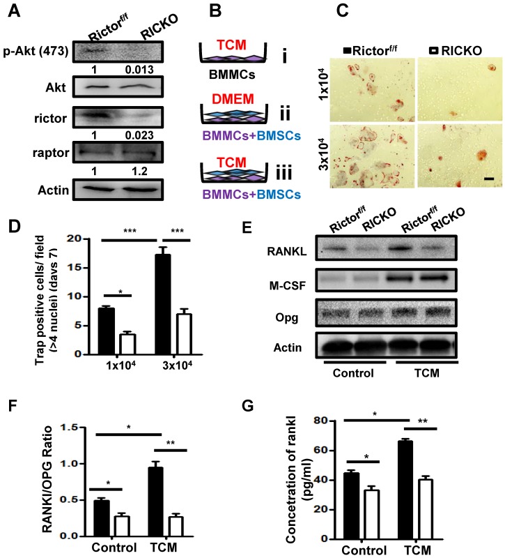
Rictor ablation in BMSCs suppresses TM40D induced osteoclastogenesis in vitro.(A) Western blot analyses of BMSCs isolated from Rictorf/f and RiCKO mice. (B) Schematic diagram of cell culture. (C) Representative TRAP staining of osteoclasts differentiated from macrophages co-cultured with BMSCs in TCM. (D) Quantification of TRAP+ cells. (E) Western blot analyses of BMSCs isolated from Rictorf/f and RiCKO mice cultured in control media or TCM.(F) Relative ratio of RANKL to OPG calculated from Western blot. (G) ELISA quantification of RANKL protein level in supernatant of BMSCs after treatment with control or conditioned medium (TCM).Scale bar=50 µm. All bar graphs show mean ± SEM. *P <0.05;**P <0.01; ***P < 0.001.

