FIG 1.
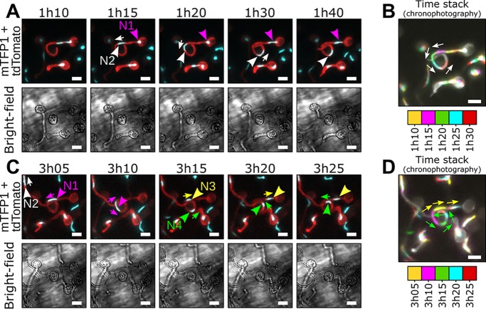
Repositioning of daughter nuclei follows division during P. palmivora cyst germination (video 1). (A to D) Time-lapse imaging of germinating cysts from a transgenic P. palmivora strain expressing cytoplasmic tdTomato as well as nucleus-localized mTFP1 (LILI-td-NT) during growth at the surface of N. benthamiana roots. (A) Following first nuclear division, daughter nucleus N1 (magenta arrowhead) migrates toward the tip of the germ tube (white arrow), while the second nucleus, N2 (white arrowhead), remains within the cyst body. (B) Chronophotographic view (time stack) of the previous sequence, showing the successive locations of nucleus N2. Time frames are color coded and overlaid. (C) Nuclear movements following N1 division. Daughter nuclei N3 (yellow arrowhead) and N4 (green arrowhead) move in opposite directions (yellow and green arrows, respectively) and distribute equally within the germ tube. (D) Chronophotographic view (time stack) of the sequence shown in panel C, showing opposite movement of nuclei N3 and N4. Bar, 10 μm.

