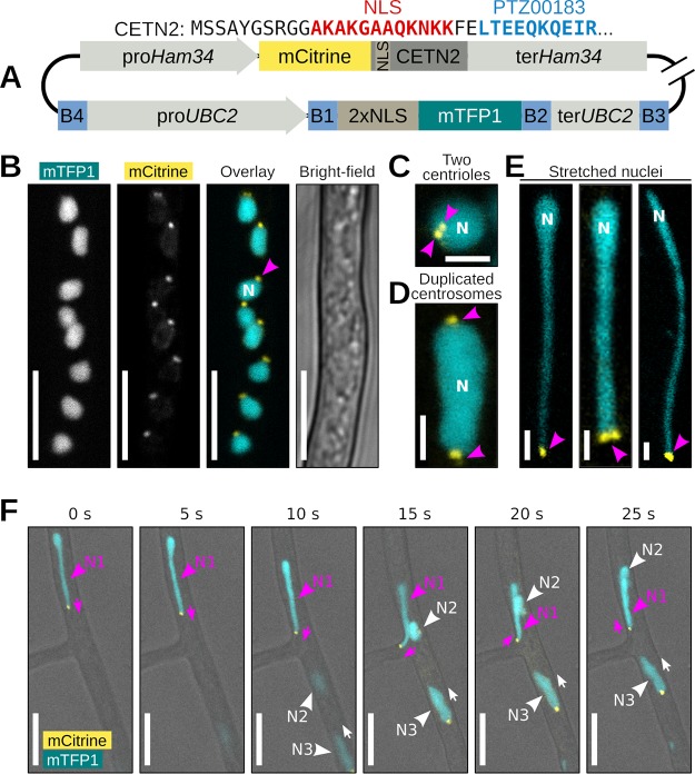
FIG 7.
Centrin2 localizes to P. palmivora nuclear stretches. (A) Schematic view of the construct used for dual labeling of nuclei and centrosomes in P. palmivora strain LILI-NT-Ce. Backbone elements are not represented. (B, video 9) Representative picture of mCitrine-labeled Centrin2 (CETN2) within P. palmivora LILI-NT-Ce hyphae. (C) CETN2 localizes in two adjacent dots at the periphery of the nucleus. (D) Centrosome duplication in dividing nuclei. (E) Representative pictures of stretched P. palmivora nuclei (up to 30 μm long), with CETN2 localizing at the tip of the stretched areas. (F) Time-lapse imaging of nuclear movements at hyphal branch point. Nucleus N1 (magenta) follows an A-type trajectory backward to the branch point, while nuclei N2 and N3 follow a P-type trajectory. CETN2 localizes at the tip of the stretched part of nucleus N1, while CETN2 localizes at the back of nuclei N2 and N3. Bar, 2 μm (C to E) or 10 μm (B and F).

