Figure 4.
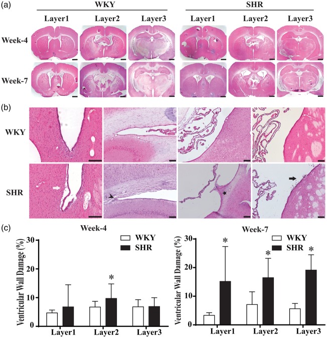
Ventricular wall damage in SHR and WKY rats at weeks 4 and 7. (a) Hematoxylin and eosin- (H & E) stained sections were examined at three different layers of ventricular system for ventricular wall damage. Scale bar = 1 mm. (b) Typical ventricle wall injury including detachment of ependymal layer (white arrow), sparse white matter (arrowhead), bulge (star) and cells shedding (black arrow) was found in SHRs at week 7. Scale bar = 50 μm (high magnification) and 100 μm (low magnification). (c) Quantification of ventricle wall damage (percentage of total perimeter). SHRs had more damage on layer 2 at week 4 and on all the three layers at week 7. Values are means ± SD; *P < 0.05 compared with WKY group; n = 6.

