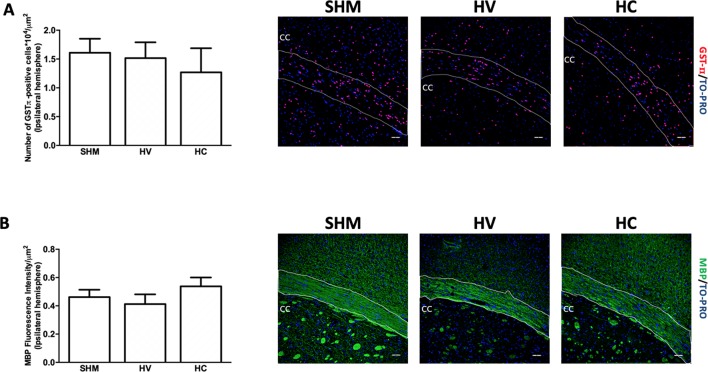
Figure 1.
Quantification of mature oligodendrocyte population and myelin staining in white matter. Representative microphotographs and graphical representation of immunohistochemical studies performed in the external capsula of the corpus callosum 30 days after a hypoxic-ischemic (HI) insult induced in P7–10 Wistar rats then receiving vehicle (HV) or cannabidiol (HC), or a similar period in control rats (SHM). (A) GST-π (mature oligodendrocyte) and (B) myelin basic protein (MBP) staining. Bars represent mean ± SEM. Scale bar: 50 μm.

