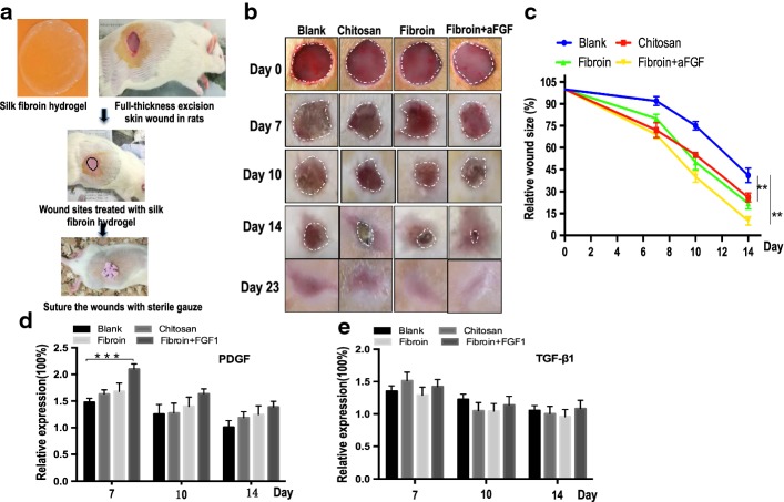
Fig. 2.
a Schematic diagram of silk fibroin hydrogel application for wound healing. The wound was formed by punching a hole through the whole skin of rats. b Extent of wound healing on days 7,10 and 14. c Measure changes in the percentages of wound closure. d, e The expression levels of PDGF and TGF-β1 in full-thickness skin defects wound using ELISA at three time points. The relative fold change was ratios between different concentration of growth factors in four operation groups and normal skins cut from rats at different time points. Asterisks indicate statistical significance based on Student’s t tests (***P < 0.001)

