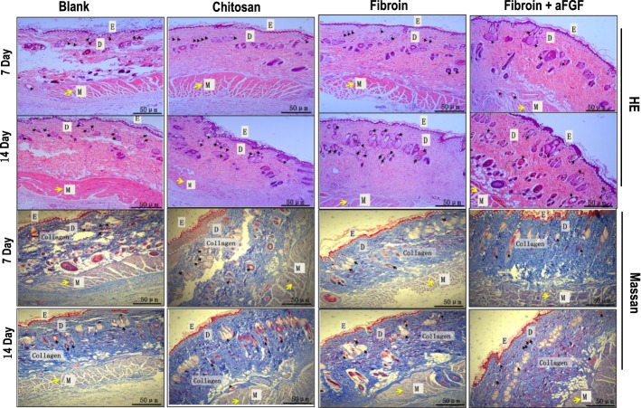
Fig. 3.
HE staining photographs (upper two panels) and MT staining images (lower two panels) of wounds. Black arrows point to the sites of epidermal differentiation into hair follicles and sebaceous glands and yellow arrows point to the sites of muscle tissue. Blue color represents collagen staining. D dermis, E epidermis, M muscle

