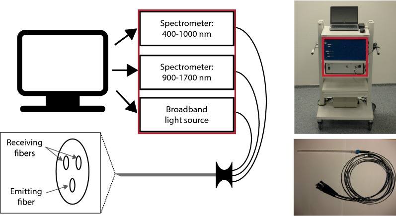
Fig. 2.
Measurement system. On the left a schematic image is shown of the system used to perform the measurements. The system consists of two spectrometers and a broadband light source, which are all controlled by a computer. Measurements are performed using a needle which includes three fibers. One that transports the light from the broadband light source to the tissue (emitting fiber) and two to transport the light from the tissue to the two spectrometers (receiving fibers). The distance between the receiving and emitting fibers is 1.29 mm. On the right, images are shown of the system as used during surgery (top image) and the needle used to perform the measurements with (bottom image)

