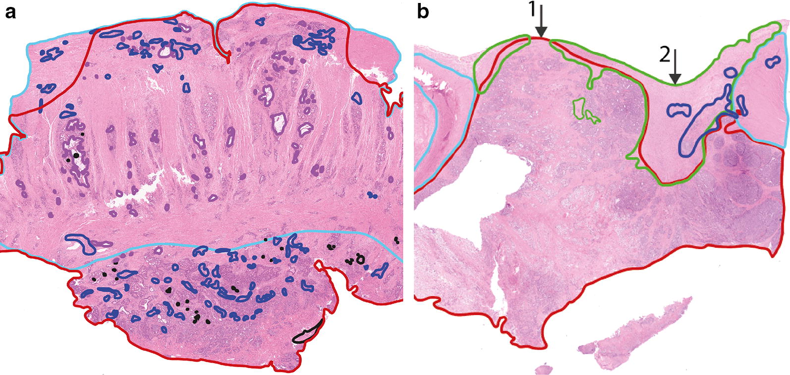
Fig. 5.

H&E slides of a measured locations with conclusive and inconclusive correlation to histopathology. H&E slides were annotated by a pathologist. Red = tumor, light blue = muscle, green = fibrosis, dark blue = inflammation. a Conclusive histopathology, with a large area of only tumor at the surface. b Inconclusive histopathology, if the measurement would have been on location 1, it would be a tumor measurement, however on location 2, less than 0.5 mm to the right it would be a fibrosis measurement. Locations with histopathology similar to b were excluded whereas locations with histopathology similar to a were used for classification
