Fig. 8.
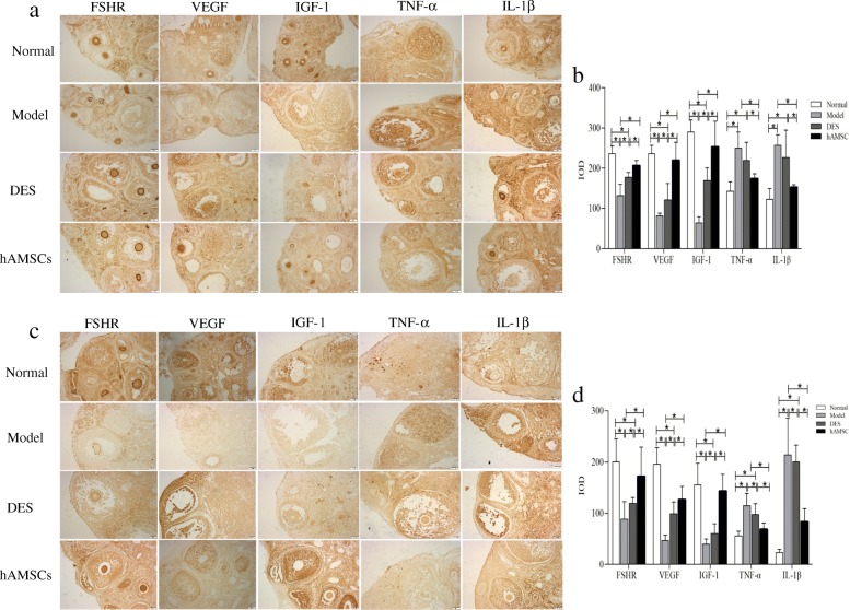
The hAMSCs improved protein expression associated with follicular development in the ovarian microenvironment of POF mice. The brown particles of positive expression were observed on FSHR, VEGF, IGF-1, TNF-α, and IL-1β proteins by immunohistochemical staining at 7 days (a) and 14 days (c) after hAMSCs transplantation. At 7 (b) and 14 days (d) after cell transplantation, the mean integral optical density value (IOD) of FSHR-, VEGF-, IGF-1-, TNF-α-, and IL-1β-positive areas was determined in ovarian tissue by Image-Pro Plus (immunohistochemical staining, × 200). Data are presented as the mean ± S ( ± s, n = 6), *P < 0.05

