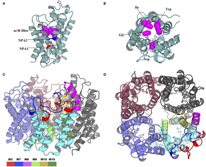
Figure 10.
Cartoon rendering of the tick AQP1 homology model and motif mapping. (A) A side-view of the modeled tick AQP1 monomer showing the position of the two Asparagine-Proline-Alanine (NPA) motifs and the aromatic-arginine (ar/R) filter. (B) A top view of the modeled tick AQP1 monomer showing the amino acid sequences that constitute the ar/R filter (chain A residues: Trp33, Gly136, Ile171, and Asp188). (C) A cartoon rendering of the tick AQP1 protein and (D). A surface view rendering of the tick AQP1 protein. Both (C,D) show the mapped tick-specific peptide motifs. Motifs M7 and M8 were located on the protein surface and in the extra-cellular domain while M9, M10, and M6 were located in the transmembrane domain. The motifs were colored as follows: M3- red, M7- blue, M8- purple, M9- light green, M10- yellow, and M16- dark green.

