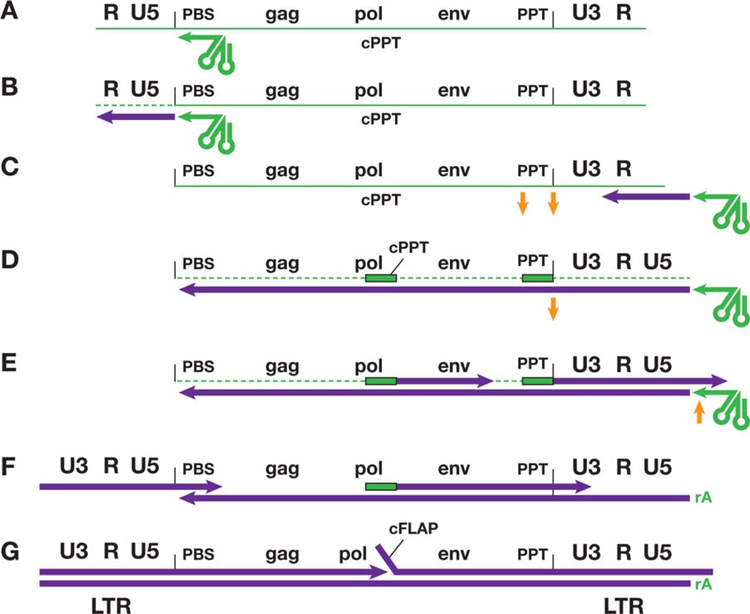
FIGURE 1.
Reverse transcription of the genome of HIV-1. This figure shows, in cartoon form, the steps that are involved in the conversion of the ssRNA genome found in virions into dsDNA. In the figure, RNA is shown in green and DNA in purple. For simplicity, the 5′ cap and the poly(A) tail, which are present on the viral genomic RNA, have been omitted. The various sequence elements in the viral genome, including the genes, are not drawn to scale. (A) The tRNA primer (green arrow on the left) is base paired to the primer-binding site (PBS). (B) RT has initiated minus-strand DNA synthesis from the tRNA primer, and has copied the U5 and R sequences at the 5′ end of the genome. This creates an RNA/DNA duplex, which allows RNase H to degrade the RNA that has been copied (dotted green line). (C) The minus-strand DNA (see text) has been transferred, using the R sequence found at both ends of the viral RNA, to the 3′ end of the viral RNA, and minus-strand DNA synthesis can resume. The HIV-1 genome has two purine-rich sequences (polypurine tracts, or PPTs, one immediately adjacent to U3; the other, the central PPT [cPPT] is in the pol gene). (D–G) The two PPTs are relatively resistant to RNase H (D) and they serve as primers for plus-strand DNA synthesis (E). Once plus-strand DNA synthesis has been initiated, RNase H removes the PPT from the plus-strand DNAs (F, G). The plus-strand that is initiated in U3 is extended until the first 18 bases to the tRNA primer are copied (see text); RNase H then removes the tRNA primer (E). It appears that the entire PPT primers are removed by RNase H; however, RNase H leaves a single riboA on the 5′ end of the minus-strand (E, F). Removing the tRNA primer sets the stage for the second-strand transfer (F). Both the plus- and minus-strand DNAs are then elongated. The plus-strand that was initiated at the U3 junction (on the left in the figure) displaces a segment of the plus-strand that was initiated from the cPPT, creating a small flap, called the central flap (cFLAP). doi:10.1128/microbiolspec.MDNA3–0027-2014.f1

