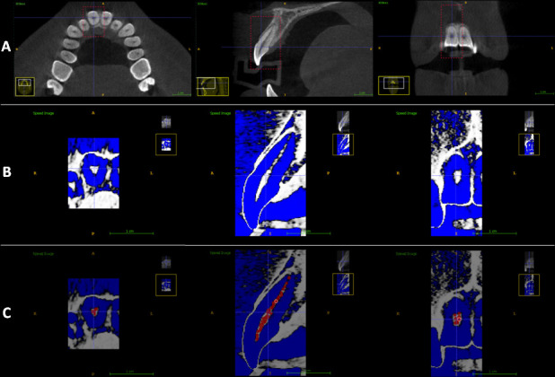
Figure 1.

Steps of the segmentation process of a right upper central incisor using CBCT imaging. (a) Selection of the region of interest (ROI) in the axial, sagittal and coronal planes. (b) After adjustment of the default density range. (c) Addition of “seeds” throughout the pulp extension in the axial, sagittal and coronal planes.
