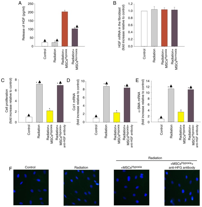
Figure 2.
Involvement of HGF in MSCsHypoxia-driven inhibition of fibroblast proliferation and radiation-induced FMT. To explore the role of the anti-fibrotic effect of MSCsHypoxia on radiation-treated fibroblast cells, a Transwell co-culture system was used. (A) ELISA measuring the release of HGF into the condition media of fibroblasts, fibroblasts treated with radiation, and fibroblasts co-cultured with MSCsHypoxia or MSCsNormoxia in the presence of radiation. (B) HGF mRNA levels in fibroblasts, fibroblasts treated with radiation, and fibroblasts co-cultured with MSCsHypoxia or MSCsNormoxia in the presence of radiation were analyzed by RT-qPCR. Each column represents the mean ± SD from three independent experiments; *P<0.05 vs. the Control; ▲P<0.05 vs. Radiation + MSCsHypoxia. In fibroblasts, fibroblasts treated with radiation, and fibroblasts co-cultured with MSCsHypoxia or MSCsHypoxia + anti-HGF antibody in the presence of radiation, (C) proliferation growth curves were determined using an MTT assay. (D and E) Col1 and α-SMA mRNA levels as analyzed by RT-qPCR. (F) Expression of α-SMA was measured using immunofluorescence staining. Each column represents the mean ± SD from three independent experiments; *P<0.05 vs. the Control; ▲P<0.05 vs. Radiation + MSCsHypoxia. HGF, hepatocyte growth factor; MSCs, mesenchymal stem cells; FMT, fibroblast-to-myofibroblast transition; Col1, type I collagen; α-SMA, α-smooth muscle actin.

