Figure 1.
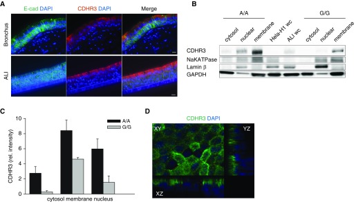
CDHR3 localizes in the apical cells of human airway epithelial cells. (A) Immunofluorescent staining of E-cadherin (E-cad; green), CDHR3 (red), and nucleus (DAPI; blue) in bronchial tissue (top) and human bronchial epithelial cell air–liquid interface (BEC-ALI) culture (bottom). (B) Immunoblot analysis of three subcellular compartments extracted from BEC-ALI cultures (n = 3) with A/A and G/G genotype: cytosol, membrane, and nuclear, stained for CDHR3 (∼100 kD). (C) Quantification of CDHR3 protein band signal intensity of subcellular compartments (n = 3). (D) Z-stack slice view of BEC-ALI culture stained for CDHR3 (green) and nucleus (DAPI). Scale bars: 10 μm. CDHR3 = cadherin related family member 3; rel. = relative; wc = whole-cell lysate.

