Figure 3.
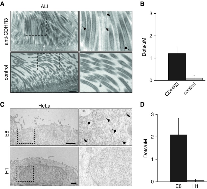
Transmission electron microscopy micrographs of immunolabeled CDHR3 in human BEC-ALI and HeLa cells. (A) BEC-ALI cultures were incubated with immunogold-labeled anti-CDHR3 monoclonal antibody (black dots, top row) or a control antibody (bottom row). Right column shows labeled CDHR3 (arrows) at a higher magnification within inset boxes. (B) Quantification of immunogold labeling in BEC-ALI cultures incubated with anti-CDHR3 or control antibody. (C) HeLa-E8 (transduced with CDHR3) and native HeLa-H1 cells were incubated with immunogold-labeled anti-CDHR3 monoclonal antibody (black dots). Right column shows labeled CDHR3 (arrows) at a higher magnification within inset boxes. (D) Quantification of immunolabeling in HeLa-H8 and HeLa-H1. Scale bars: 1 μm.

