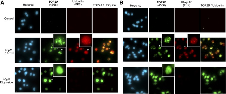
Fig. 3.
PR-619–induced TOP2 complexes are unevenly distributed in the nucleus. Cells were treated with PR-619, etoposide, or solvent control (DMSO) for 2 hours and TOP2 complexes were detected using the TARDIS assay. Immunofluorescence was performed using anti-TOP2A (A) or anti TOP2B (B) antibodies and anti-ubiquitin (FK2). Images were acquired using a 40X objective and are extended focus widefield images. Enlarged nuclei indicated by asterisk; scale bar, 10 μm.

