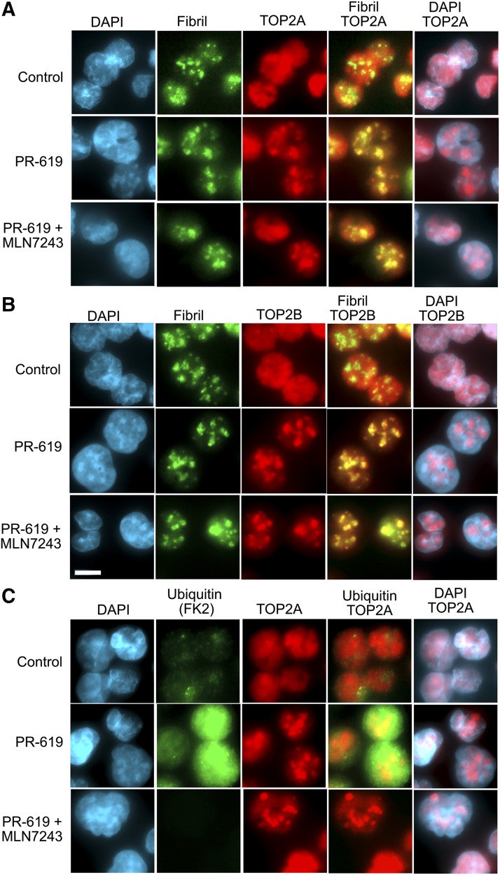
Fig. 4.
PR-619 drives nucleolar localization of TOP2A and TOP2B. K562 cells were treated with PR-619 (80 µM), and where indicated were pretreated with the ubiquitin-activating enzyme inhibitor MLN7243 (10 μM, 2 hours), fixed with paraformaldehyde, and analyzed by immunofluorescence for TOP2A (A) or TOP2B (B) and the nucleolar marker fibrillarin. (C) K562 cells were treated as in (A and B) and stained for ubiquitin (FK2) and TOP2A. Images are shown as extended focus projections using 0.1 μm z-steps and representative images obtained from several fields of cells from duplicate slides; scale bar, 10 μm.

