Fig. 8.
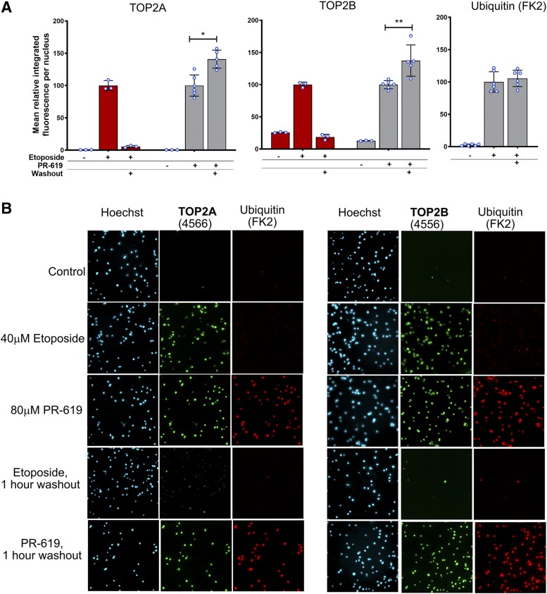
PR-619–induced TOP2 complexes persist after drug washout. K562 cells were incubated in the presence of etoposide (40 µM), PR-619 (80 µM), or solvent (DMSO) for 2 hours. Cells were either collected immediately or drug was washed out and cells were replated and incubated for a further 1 hour before collection for TARDIS analysis. (A) Quantification of TARDIS data obtained using anti-TOP2A (4566), anti-TOP2B (4556), and anti-ubiquitin (FK2) antibodies. Median-integrated fluorescence per nucleus values were normalized to the mean of the medians obtained with each drug directly after 2-hour incubation (i.e., without washout). Data are shown as mean ± S.D. values, and values from individual replicas are indicated as blue-lined circles. Statistical analysis, the normalized mean integrated fluorescence for PR-619 before and after washout were compared by unpaired t test. (B) Representative images from TARDIS slides used to produce part A.

