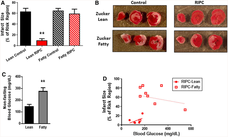
Fig. 2.
a Protocol 1: infarct size, expressed as a % of the myocardium at risk (mean ± SEM), for Zucker lean and Zucker fatty rats randomized to receive remote ischemic preconditioning (RIPC) or a time-matched control period. **p < 0.01 versus the Zucker lean control group, b Protocol 1: images of heart slices obtained from one control and one RIPC-treated rat from the Zucker lean and Zucker fatty cohorts. Heart slices were incubated in triphenyltetrazolium chloride; using this method, viable myocardium stains red while areas of necrosis remain unstained, and thus appear pale, c Protocol 1: non-fasting blood glucose concentration (mg/dL; mean ± SEM) for Zucker lean and Zucker fatty rats. **p < 0.01 versus Zucker lean rats, d Protocol 1: infarct size (expressed as a % of the myocardium) plotted as a function of non-fasting blood glucose concentration (mg/dL) for Zucker lean and Zucker fatty rats that underwent RIPC

