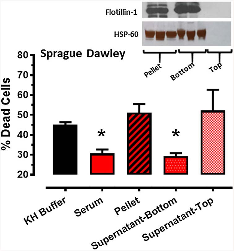
Fig. 3.
Protocol 2: effect of Krebs–Henseleit (KH) buffer, serum and serum fractions (pellet, supernatant-bottom, supernatant-top), obtained from Sprague–Dawley rats following the standard RIPC stimulus, on % cell death (mean ± SEM) of HL-1 cells subjected to 2.5 h of hypoxia and 24 h of reoxygenation. *p < 0.05 versus KH buffer. Inset: expression of flotillin 1 and HSP-60 in the pellet, supernatant-top and supernatant bottom serum fractions from Sprague–Dawley rats following RIPC

