Fig. 1.
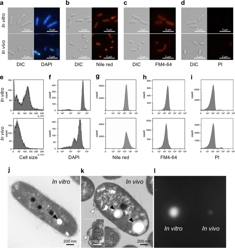
Bacterial cell morphology of in vivo and in vitro Burkholderia symbiont cells. a–d Differential interference contrast (left) and fluorescence microscopy (right) images of in vitro and in vivo bacteria stained with DAPI a, Nile red b, FM4-64 c, and PI d. e–i Flow cytometry analysis of in vivo and in vitro symbiont cells measuring cell size by light scatter e, DNA content by DAPI staining f, PHA accumulation by Nile red staining g, and membrane area by FM4-64 staining h, membrane permeability by PI staining i. j, k Transmission electron microscopy images of an in vitro j and an in vivo k symbiont cell. Filled arrows indicate PHA granules and open arrows shows a membrane bleb. l Motility test of in vitro (left) and in vivo (right) cells (see also Supplementary Fig. S3)

