
Fig. 6.
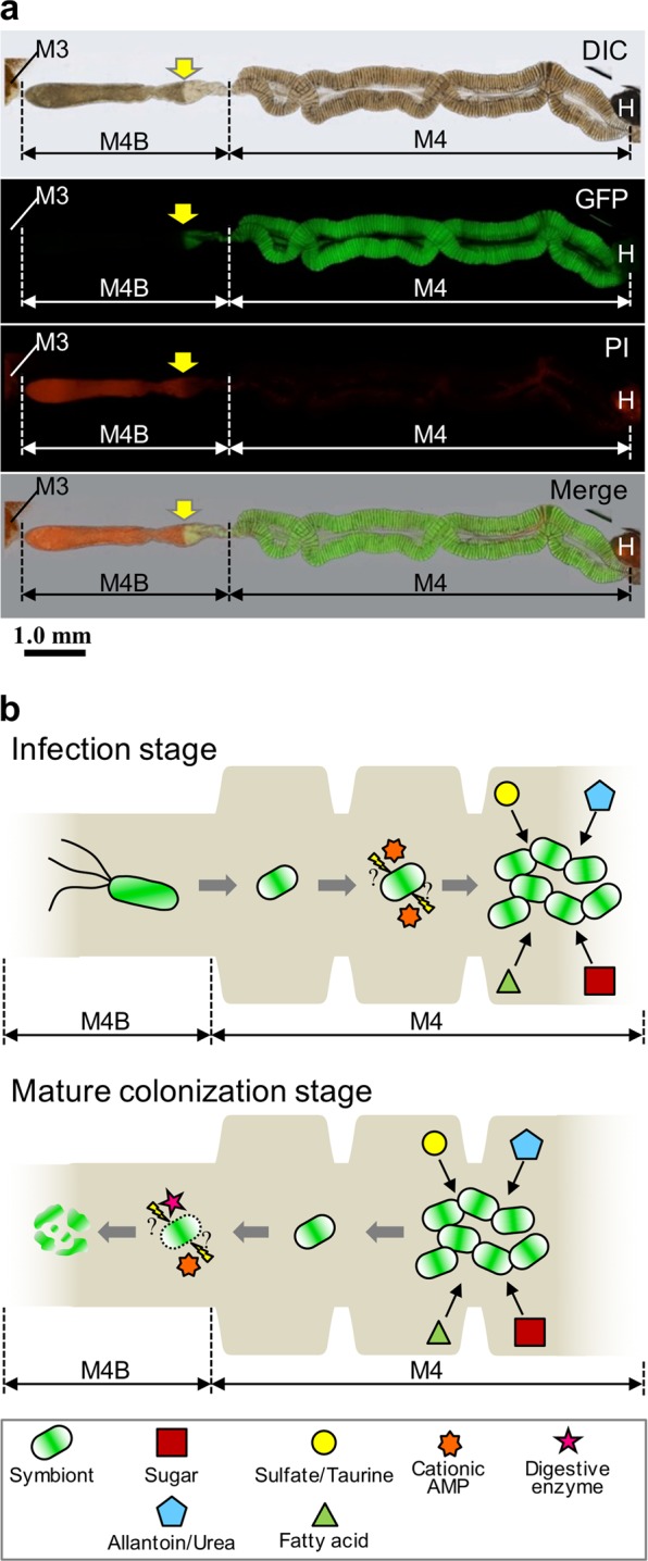
A hypothetical model for the life cycle of the Burkholderia symbiont in the midgut of R. pedestris. a The PI staining in the midgut of 5th instar nymph infected with the strain RPE225 (a GFP-expressing mutant). DIC, GFP fluorescence, PI fluorescence, and merged images are shown. Arrows indicate the border of GFP and PI fluorescent signals. M3, midgut 3rd section; M4, midgut 4th section (crypts); M4B, M4 bulb; H, hindgut. A red auto-fluorescence was observed in M3 and H. b Graphical summary of Burkholderia features and midgut functions of R. pedestris. (Upper) During the infection stage of Burkholderia symbiont at the insect midgut, the Burkholderia symbiont cells lose flagellar motility and modify their envelope under the influence of host stress factors such as cationic AMPs (e.g. CCRs), they proliferate by metabolizing host waste materials, such as sulfate and allantoin. (Lower) In the mature colonization stage, these symbiont cells are digested in the M4B section mediated by host factors such as cathepsin proteases and CCRs, and the host absorbs nutrients derived from whole-bacterial cells