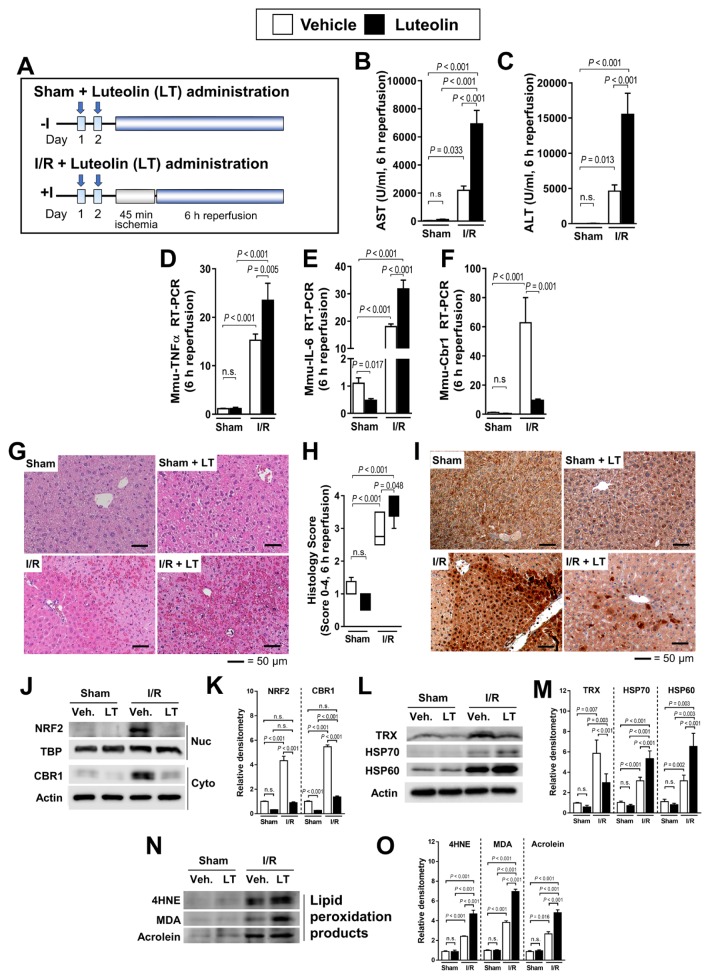
Fig. 4. Administration of the CBR1 reducer LT increases hepatic ischemia–reperfusion injury and hepatic cell death.
(A) Schematic outline of LT administration of mice. Serum measurement of (B) AST and (C) ALT. Relative expression of (D) murine TNFα, (E) IL-6, and (F) Cbr1 measured by qRT-PCR. Expression levels are normalized to Gapdh. (G) H&E staining of liver injury and (H) histology score of LT-treated mice. (I) Immunohistochemistry of CBR1. (J and K) Protein expression of Nrf2 and CBR1 and relative density. (L and M) Western blot analysis of oxidative stress markers (TRX, HSP70, and HSP60) and relative density. (N and O) Western blot of lipid peroxidation products (4HNE, MDA, and Acrolein) and relative density. Protein levels were normalized to Actin, except for Nrf2 which was normalized to TBP. The levels of other compounds were normalized to matching control group. Veh., vehicle; White bar, vehicle-treated; Black bar, LT-treated. Comparison is indicated by a line and P value. n.s., not significant. n = 6–12 per group.

