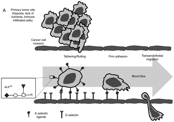
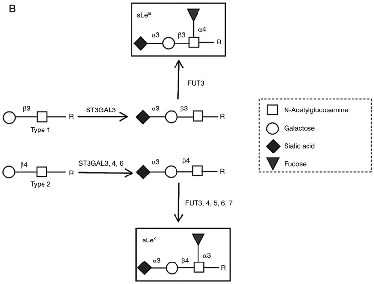
Figure 1.
Schematic representation of the multistep metastatic process in cancer and main structures involved. (A) Contribution of sLex/sLea antigens in facilitating cell adhesion to E-selectins. There are four main steps in the extravasation process, including tethering and rolling, integrin activation, firm adhesion and transendothelial migration. Metastatic dissemination is facilitated by interactions between tumour cells and endothelium in distant tissues. These interactions are mediated by E-selectins expressed by activated endothelial cells and the corresponding E-selectin ligands expressed on the cell surface of cancer cells. These ligands are prototypically the sLex and sLea antigens, displayed on cell surface proteins or lipid scaffolds. (B) Structure and schematic representation of the biosynthesis of sialyl Lewis antigens. sLea and sLex antigens are sialofucosylated isomer tetrasaccharides, derived from type 1 or type 2 sugar chains, respectively, attached to N- or O-glycan residues. The two types of structures are sialylated by the action of the indicated α2,3-sialyltransferases and successively fucosylated by the action of the FUT3 (in type 1 chains), and FUT3, 4, 5, 6 or 7 (in type 2 chains). FUT, fucosyltransferase; sLea/x, sialyl Lewis a/x.


