Figure 2.
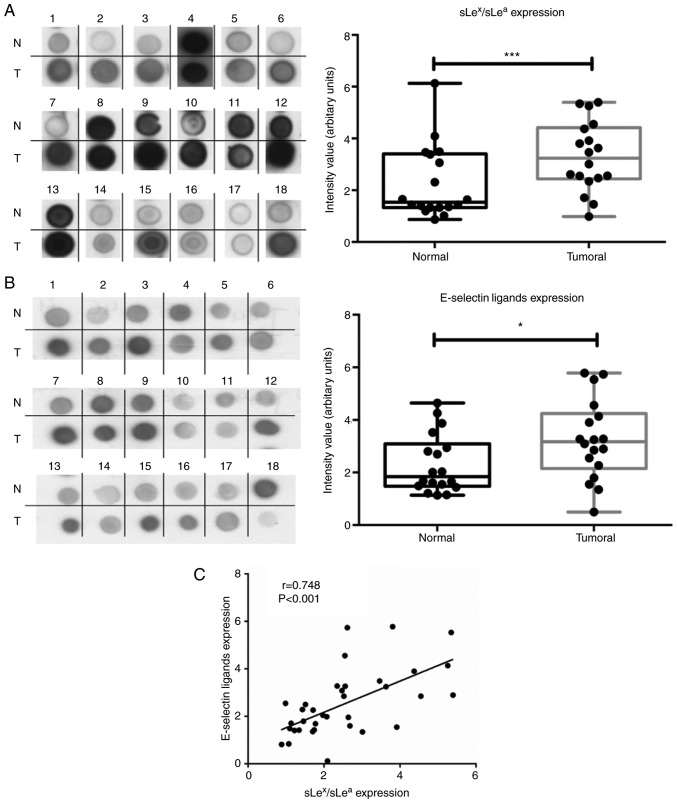
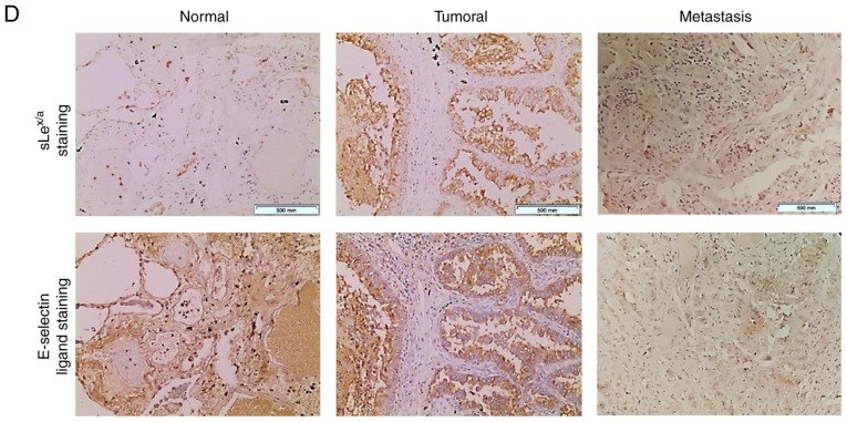
NSCLC has increased anti-sLex/sLea mAb and E-selectin reactivity. (A) Dot blot analysis of anti-sLex/sLea mAb reactivity in matched normal and tumour proteins. Total lysates of N or T tissues were spotted on nitrocellulose membrane and stained with HECA-452 mAb. Left panel, representative dot blot analysis of the HECA-452 mAb reactivity. Right, the intensity of each dot blot spot, determined using ImageJ 1.48v software and expressed as arbitrary units. Box-and-whisker plots represent median, and lower and upper quartile values (boxes), ranges and all values for each group (black dots). (B) Dot blot analysis of E-selectin reactivity in matched normal and tumour proteins. Total lysates of N or T tissues were spotted on nitrocellulose membrane and stained with E-Ig. Left, representative dot blot analysis of E-Ig reactivity. Right, the intensity of each dot blot spot expressed as arbitrary units. Box-and-whisker plots represent median values, and lower and upper quartiles (boxes), ranges and all values for each group (black dots). *P<0.05, ***P<0.001. (C) Correlation between anti-sLex/sLea mAb and E-Ig staining intensity in tumour tissue. Correlation was analysed using Spearman's correlation coefficient. NSCLC has increased anti-sLex/sLea mAb and E-selectin reactivity. (D) Anti-sLex/sLea mAb and E-Ig reactivity in paraffin-embedded normal, tumour and metastasis sections. Sequential sections from representative normal, non-small cell lung cancer and bone metastasis tissues were stained with HECA-452 mAb (top) and E-Ig chimera (bottom) via immunohistochemistry. Nuclei stained with haematoxylin. Magnification, ×10. E-Ig, mouse E-selectin-human Fc Ig chimera; mAb, monoclonal antibody; N, normal; sLea/x, sialyl Lewis a/x; T, tumour.


