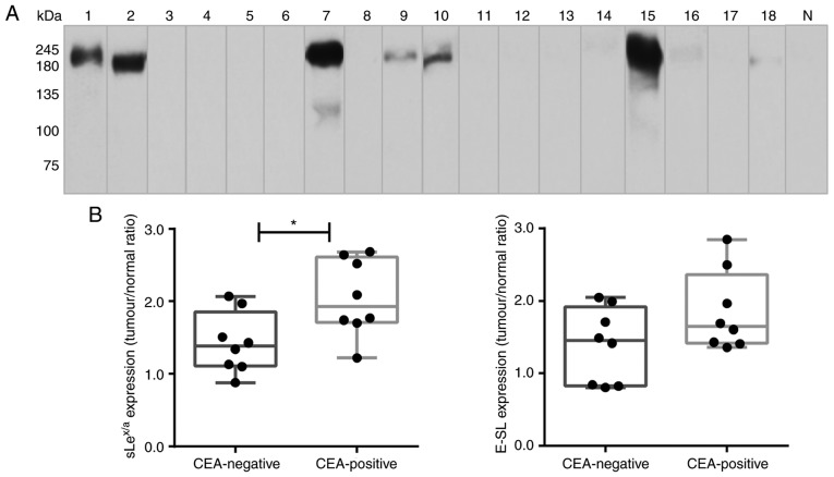
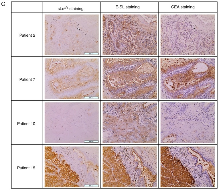
Figure 4.
CEA is expressed in patients with NSCLC. (A) Western blot analysis of CEA glycoprotein in tumour lysates. Protein (20 µg) obtained from NSCLC patients' tissues were ran in reduced SDS-PAGE gels and blotted with anti-CEA mAb. The numbers above each lane represent the patient number. The lane N corresponds to a representative normal tissue lysate from a patient with NSCLC (patient number 7), showing negative anti-CEA reactivity. This figure shows a blot with tracks from samples analysed separately. (B) sLex/sLea and E-selectin ligands expression in CEA-negative and CEA-positive patients. The tumour/normal ratios of sLex/sLea expression (left) and E-selectin ligands expression (right) were calculated and separated for CEA-negative and CEA-positive patients. Box-and-whisker plots represent median values, and upper and lower quartiles (boxes), ranges and all values for each group (black dots). *P<0.05. (C) sLex/sLea, E-selectin ligands and CEA have overlapping staining profiles in NSCLC tissues. Sequential paraffin-embedded NSCLC tissue sections from patients 2, 7, 10 and 15 were stained with HECA-452 mAb (left), E-Ig chimera (middle), and anti-CEA mAb (right) via immunohistochemistry. Nuclei were stained with haematoxylin. Magnification, ×10. CEA, carcinoembryonic antigen; E-Ig, mouse E-selectin-human Fc Ig chimera; E-SL, E-selectin; mAb, monoclonal antibody; N, normal; NSCLC, non-small cell lung cancer; sLea/x, sialyl Lewis a/x.


