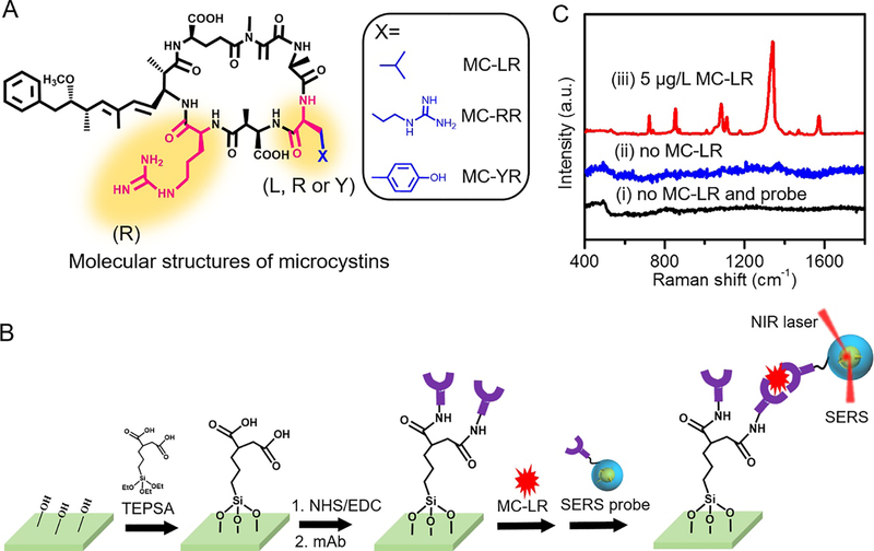
Figure 2. Detection of MC-LR.
(A) Molecular structures of microcystin-LR, microcystin-RR and microcystin-YR. (B) Schematic illustration of fabrication of the SERS immunoassay platform and its operation principle for microcystin-LR detection. (C) Representative SERS spectra obtained from the developed assay – (i) in the absence of both MC-LR analyte and SERS probes, (ii) in the presence of SERS probes and absence of MC-LR analyte and (iii) in the presence of both 5 μg/L MC-LR analytes and SERS probes.

