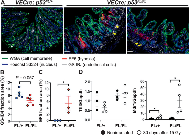
FIG. 3.
Delayed radiation-induced intestinal injury is characterized by microvessel damage and tissue hypoxia. Panel A: Representative tissue sections of one VECre; p53FL/+ mouse and two VECre; p53FL/FL mice at day 30 after 15 Gy TAI. The small intestines of irradiated VECre; p53FL/FL mice showed a decrease in GS-IB4+ endothelial cells within the lamina propria of the villi. In addition, the small intestines of irradiated VECre; p53FL/FL mice, but not the VECre; p53FL/+ mouse, exhibited areas of hypoxia as shown by EF5 staining. Note that in irradiated VECre; p53FL/FL mice, even in regions with loss of microvessels in the villi and associated hypoxia, the adjacent crypts (yellow arrows) are largely intact. Scale bar: 100 μm. Panels B and C: Quantification of the area stained positive for GS-IB4 or EF5 per high-power field 30 days after 15 Gy TAI. P value was calculated using Student’s t test. Each dot represents one mouse. Panel D: The expression of hypoxia-responsive genes Tff3 and Mdr1 (Abcb1) in intestinal epithelial cells from unirradiated mice or at day 30 after 15 Gy TAI. *P < 0.05 (two-way ANOVA with Bonferroni post hoc test).

