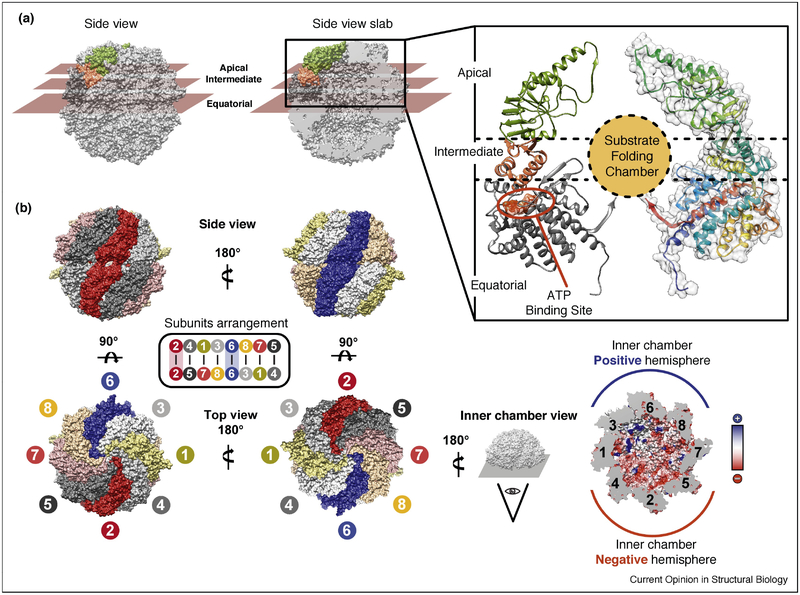
Figure 1: TRiC Architecture and Subunit Arrangement.
(A) Closed TRiC structure with the three domains in a single subunit highlighted: equatorial domain (dark gray) responsible for binding ATP, apical domain (green) responsible for binding substrate and forming the built in lid, and hinge domain (salmon) responsible for relaying ATP changes into movements of the apical domain. Domain distribution indicated by planar sectioning, and also indicated on ribbon diagram of single subunit. (B) Subunit arrangement of TRiC, rings stacked back to back with CCT2 (red) and CCT6 (blue) stacked on top of each other. (B, lower right) Charge asymmetry in the interior of the closed chamber: one hemisphere (blue) is lined with positively charged side chains (+)and the other hemisphere (red) lined with negatively charged side chains (−). Inner chamber electrostatic potential also displayed. (PDB structures used: 4V94)

